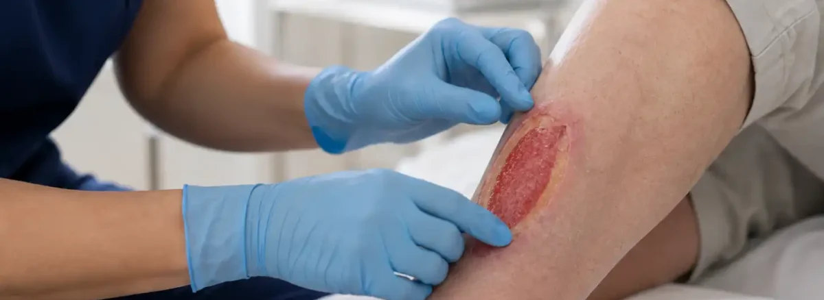
What color should a healing wound be? Somewhere between firm red and pale pink, and shading further toward pink as it closes. That is the short answer, and it is true of almost every open wound on the body.
The longer answer is that almost nobody is looking at a wound that is only one color, and the color people fix on is usually the one that alarms them most. This guide covers the shades a wound bed can show, the mix-up that sends most people looking at the wrong part of the wound entirely, why yellow is far less frightening than it looks, and how to read a color as a direction of travel instead of a verdict.
On this page
- What color should a healing wound be?
- Red inside the wound is not red around it
- Almost no wound is a single color
- What each color in the wound bed means
- Pink is the shade to hunt for
- Yellow is not automatically infection
- Black, purple and green
- How to look at it properly
- What to avoid
- Living with a wound you check every day
- When to get it seen
- Questions about wound color
- The takeaway
What color should a healing wound be?
A wound that is repairing itself well shows a firm, even red across its base, with pale pink creeping in from the edges. The red is granulation tissue, the new material the body builds to fill a hole in itself. It looks slightly bumpy, sits moist without being wet, and tends to bleed if it is rubbed. The pink is newer still, the thin sheet of skin cells moving inward to roof the whole thing over.
StatPearls, published through the National Library of Medicine, describes healthy granulation in its chapter on wound assessment as beefy red tissue that bleeds and signals repair underway, and sets it against pale yellow tissue that does not bleed. That contrast (red and bleeding against yellow and inert) is the one distinction most of this page rests on.
Anything else you can see is a variation on those two things or a sign that something is holding the wound back. None of the variations is a diagnosis on its own, and the point of learning them is not so you can grade your own wound. It is so you can describe it accurately to the person who can.
Red inside the wound is not red around it
This trips up more people than any other part of reading a wound, and it accounts for a good share of the calls that come in worried about redness.
Red in the wound bed (the open surface at the bottom) is what you want. Red on the intact skin surrounding the wound is a different signal entirely, and the two get read as the same thing because the word is the same.
A narrow band of pink or light red hugging the wound edge, perhaps a quarter of an inch wide, is ordinary repair activity and shows up on wounds that are doing well. What concerns a clinician is redness that pushes outward across healthy skin, feels warmer than the limb elsewhere, and covers more ground on Thursday than it did on Monday. Swelling, climbing pain or streaks running away from the wound shift the picture again, and how to tell if a wound is infected works through each signal in turn.
On brown and black skin, that spreading redness may not read as red at all. It can appear as an area that looks deeper in tone, purplish or simply different from the skin beside it, and it is often easier to feel than to see. Warmth, firmness and puffiness under the fingertips carry more information there than color does, which is a reason to touch the skin around a wound and not only photograph it.
So when you describe a wound to a nurse and use the word red, say where. Inside the wound or on the skin outside it: that one word changes the whole meaning of the sentence.
Almost no wound is a single color
Ask what color a wound is and you are already asking a question the wound cannot answer. Open a dressing on a leg ulcer that has been there four months and you will typically find red in one part, yellow film over another, and a rim of pink at the edge, all at once.
Which is why clinical notes record proportions. A wound gets written up as roughly seventy percent granulation and thirty percent slough, or half and half, and those percentages are re-estimated at each visit. Nobody is trying to be precise to the square millimeter. The number exists so the next person can tell which way the wound moved.
The direction the proportions are moving tells you far more than any single day’s color. A wound bed that was two-thirds yellow last month and is two-thirds red now is a wound heading the right way, even if the yellow that remains still bothers you. A bed that was mostly red and is filling with yellow has changed course, and that deserves a conversation whether or not anything hurts.
Snapshots mislead because wounds move slowly. Looking every day is close to useless for judging progress and reliably makes people feel nothing is happening. A week apart, in the same light, is where change becomes visible.
What each color in the wound bed means
Six shades cover nearly everything a person is likely to see. Each one describes a kind of tissue, and each kind of tissue has different implications for what happens next.
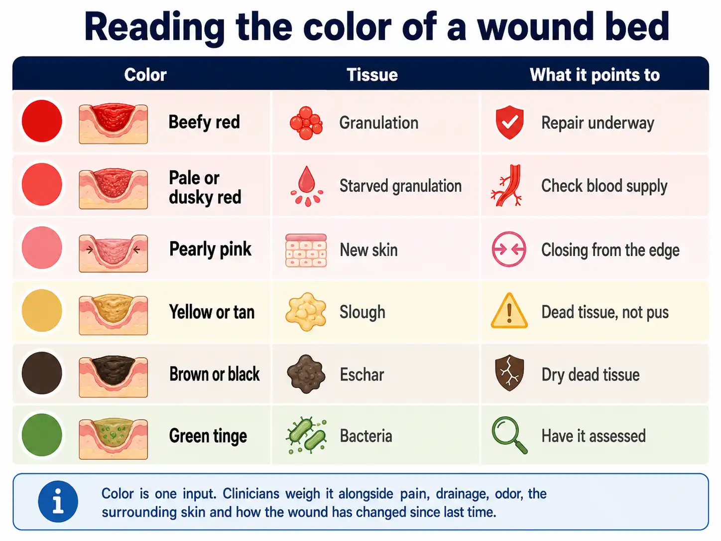
The row people ask least about is the second one, and it is the one with the most consequence. Granulation that has gone pale, gray-red or dull plum is not healthy tissue in poor lighting. It is often tissue that is not receiving enough blood to do its job, and no dressing will fix that.
A wound on the lower leg or foot whose bed looks washed out, feels cold to the touch, and hurts more when the leg is raised is a wound where circulation needs testing before anything else is decided. That combination points toward the pattern described in arterial ulcers, where the limiting factor is arterial supply, not wound care technique.
There is also such a thing as too much red. Granulation occasionally overgrows, rising above the level of the surrounding skin as a shiny, dark red, spongy mound that bleeds at the lightest touch. It is sometimes called proud flesh. It looks like enthusiastic healing and does the opposite, because new skin cannot climb a hill: the sheet of cells that should be roofing the wound stalls at the base of the mound. It is straightforward to treat once someone identifies it, and it is a good reason not to assume red always equals progress.
Pink is the shade to hunt for
If you are going to watch one color, watch pink. It is the color of a wound actually getting smaller, and it is quieter and easier to miss than any of the alarming ones.
New skin arrives as a pale, faintly shiny pink or pearl-colored band at the wound margin, thinner and flatter than the granulation beside it. It advances inward a millimeter or two at a time. On shallow wounds it can also surface in the middle of the bed as small pink islands, seeded from the remnants of hair follicles and sweat glands that survived at the base. Those islands spread outward and eventually join up with the edge.
Islands appearing in the middle of a wound bed genuinely is good news, and it is one of the few things you can see at home that reliably means the wound is winning.
Two things stop pink in its tracks and both are correctable. New skin cells cannot cross dead tissue, so slough or eschar sitting between the edge and the center holds them at the boundary. And they cannot survive a wound bed that has dried out, which is the reason dressings are designed to keep a wound moist and the reason letting a wound bed scab hard is usually the slower route. That fresh pink skin also stays fragile for a long while after it arrives, a point covered in when to stop covering an open wound.
Yellow is not automatically infection
Yellow causes more unnecessary worry than every other color combined, because people see yellow and think pus.
Most yellow in a wound bed is slough: soft dead tissue, yellow through tan through gray, stringy or filmy, stuck down to the surface underneath. It is a mixture of tissue that has died, protein and spent immune cells. It accumulates on wounds of long standing, and it turns up on plenty that carry no infection at all.
Pus behaves differently. It is liquid. It collects, it runs, and it lifts off when the wound is rinsed, leaving the bed beneath it visible. Slough stays put when irrigated and has to be taken off deliberately. If rinsing the wound clears the yellow away, you were looking at drainage; if the yellow is still there afterward, it is attached tissue.
Two further sources of yellow are worth ruling out before anyone worries. Gel-forming dressings leave a soft yellowish residue on the wound as they absorb fluid, and it can look startlingly like slough until it is rinsed off. And a stitched surgical incision often develops a thin yellow film along the line within a few days, which is fibrin laid down as part of normal closure.
Genuine slough does need clearing, because new skin will not grow across it and it feeds bacteria. Removing it is called debridement, and the gentler versions can happen under a dressing over days, or through an ointment applied once a day that breaks the tissue down chemically. Those two approaches are set out in autolytic debridement and enzymatic debridement. Sharper methods exist where speed matters. Which one suits a given wound depends on the blood supply, the amount of tissue and how quickly it needs to be gone.
Black, purple and green
The darker end of the range carries the most weight, and the three colors there mean three unrelated things.
Black or dark brown in the wound
This is eschar, dead tissue that has dried into a hard leathery cap. It hides whatever lies beneath it, so no one can judge the depth of the wound while it is in place. Some eschar is removed promptly. Dry, hard eschar on a heel, on a limb with poor circulation, is sometimes left deliberately intact as a biological cover, which surprises families who expect it to be cut away. That reasoning is explained in unstageable pressure ulcers. Black tissue on a toe or forefoot accompanied by pain, coldness or a rapid change is a different matter and needs assessment quickly.
Purple or deep maroon under unbroken skin
A purple, maroon or boggy patch on skin that has not opened, over a heel, hip, tailbone or the back of the head, can indicate damage that started in the tissue underneath and has not surfaced yet. It is easily mistaken for a bruise. What separates the two is usually the location and the story, and the response is to relieve pressure on that spot immediately and have it looked at. See deep tissue pressure injury for how those are recognized.
A green or blue-green tinge
Green staining on the wound or on the dressing pad, often with a sweetish smell, is loosely associated with one particular group of bacteria. It is worth reporting, though it is not proof of an infection needing treatment on its own. Bacteria live on most open wounds without causing harm, and what turns colonization into infection is the wound getting worse: more pain, more fluid, spreading redness, a stall in progress or feeling unwell in yourself.
How to look at it properly
Most of the value here comes from a handful of habits, none of which take long.
Judge the color after cleaning, never at the moment the dressing lifts off. Old fluid, gel residue and dressing fibers sit on the surface and color everything you are trying to read. Rinse the wound the way you were shown, give it a moment, then look.
Look in daylight. Bathroom bulbs push a wound yellow or gray, and a camera flash flattens the bed into a uniform pinkish wash that hides exactly the variation you want to see. Near a window with the flash off is a genuinely different picture from the one you get standing under a ceiling light at ten at night.
Photograph it on the same day each week, from the same distance and angle, with something for scale beside it. Comparing two photographs a week apart shows change that daily inspection never will. Keeping a short written note alongside them (roughly how much of the bed was red, how much was yellow) makes the comparison sharper still, and it makes an appointment considerably more productive.
Report the change, not the color. “It was mostly red two weeks ago and it is mostly yellow now” is a far more useful sentence to a clinician than “it looks yellow.” The routine side of dressing changes, cleaning and supplies is covered in wound care at home.
What to avoid
Do not scrub or scrape at the wound bed to clear yellow tissue. Slough is attached to living tissue underneath, and rubbing it takes granulation with it while leaving most of the slough in place. The wound goes backward and it hurts. The same applies to picking at the edge of eschar to lift it.
Keep hydrogen peroxide, undiluted antiseptics, alcohol and household disinfectants off the wound. They kill the fragile cells building the new tissue as readily as they kill bacteria, and a wound bed treated that way tends to go pale and stop advancing.
Do not let a wound bed dry out in the belief that air will help it. A dried surface stops the sheet of new skin cells from moving across, which is the slowest way to close a wound that could have closed faster.
Avoid drawing conclusions from a single glance under poor light, and avoid the opposite trap of accepting a wound that has looked identical for weeks. Unchanging red is not the same as healing red. A wound that has held the same appearance for a month without shrinking has stalled, and a stalled wound needs someone to work out why.
Living with a wound you check every day
There is a particular kind of exhaustion that comes with watching a wound, and it rarely gets acknowledged.
Every dressing change becomes a small verdict. People start reading meaning into shifts that are nothing more than lighting or the angle of the leg, and a bad-looking day can flatten a whole week. Where a family member does the dressing, the burden lands on them twice: once doing it and once deciding whether what they saw was worse.
Two habits take most of the weight off. Move the assessment to a weekly rhythm and let the daily changes just be dressing changes. And let the photographs carry the memory, because human recall of color across two weeks of Tuesdays is unreliable and always tilts toward whatever the last look happened to show.
It also helps to know that a wound getting temporarily messier is expected in some treatments. Dead tissue softening and lifting makes a wound bed look wetter, yellower and generally worse for a stretch before it looks better, and being told that in advance is the difference between a normal phase and two weeks of quiet dread. If nobody has explained what your wound is likely to look like next, ask. It is a reasonable question and the answer costs a minute.
When to get it seen
Arrange a review when the balance of the bed shifts away from red, when yellow or black tissue appears where there was none, when the bed turns pale or dull, or when the wound simply looks the same as it did a month ago.
Seek same-day attention for a raised temperature or shaking chills alongside a wound, redness traveling outward across the skin, pain that is climbing quickly, a foot or toe turning dark, or feeling abruptly unwell. Those suggest something that has moved past the wound itself.
Anyone with diabetes, reduced sensation in the feet or known circulation problems should set the threshold lower and have foot wounds looked at early, since the usual warning of pain may be absent. A wound open longer than a month with no measurable progress justifies specialist assessment on its own, whatever color it happens to be.
Questions about wound color
Is a red wound bed always a good sign?
Usually, when the red is firm and even and the wound is shrinking. Red that has gone pale, dusky or gray suggests the tissue is short of blood supply, and red that has mounded up above skin level can block new skin from spreading across. Color is read together with size, depth and progress over time.
What does pink tissue in a wound mean?
Pale, slightly shiny pink at the wound margin or as small islands in the bed is new skin forming, and it is the clearest visible sign a wound is closing. It is thinner and flatter than the red granulation around it and stays delicate for weeks after the wound looks shut.
Should I be worried about a dark red or purple ring around my wound?
A narrow band of color right at the edge is often ordinary repair. Concern rises when the area widens day by day, feels warm or firm, swells, or comes with increasing pain. On darker skin it may look deeper in tone instead of red, so compare it by touch with the skin further away and report anything that is growing.
Can a wound change color overnight?
The tissue itself changes slowly, but the appearance can shift quickly for uninteresting reasons: a different dressing, gel residue, dried blood, the lighting in the room. A genuine overnight change accompanied by pain, swelling, spreading redness or feeling unwell is the version that needs prompt attention.
Does a scab mean the wound is healing well?
A scab means the surface dried and clotted, which is not quite the same as healing efficiently. Small everyday cuts do fine under one. On larger or long-standing wounds a hard dry crust slows the new skin trying to travel underneath it, which is why wound clinics keep those beds moist under a dressing.
How quickly should the color of a wound improve?
There is no fixed timetable, and it depends on the cause, the blood supply, infection, pressure on the site and the person’s general health. A practical benchmark used in wound care is that a wound should be measurably smaller after four weeks of appropriate treatment. If neither the size nor the makeup of the bed has moved in that time, the reason for the delay is the thing to investigate.
The takeaway
So what color should a healing wound be? Firm red across the bed, turning pink from the edges inward as it closes. Pale or dusky red points at blood supply. Yellow is usually dead tissue and not pus. Black hides the depth. Purple under unbroken skin is its own problem, and green belongs in a report to your care team.
Two ideas do most of the work. Be clear about whether you are describing the wound bed or the skin around it, because red means opposite things in those two places. And judge the wound on where it is heading over a week, in daylight, after cleaning. A single glance at a single color is the least reliable information a wound will ever give you.
Not sure what you are looking at?
An assessment can identify the tissue in the wound bed, check whether blood supply is limiting progress, and set out what needs clearing before the wound can close. Bring your photographs if you have been taking them.
Wounds that have stopped making progress are handled through chronic wound treatment, where identifying what is holding the wound back comes before any change of dressing.
This article is general education and does not replace an examination of the wound itself. Color is interpreted alongside findings that cannot be judged from a photograph, and any wound that is changing appearance, failing to shrink, or accompanied by fever or spreading redness should be assessed by a healthcare professional.