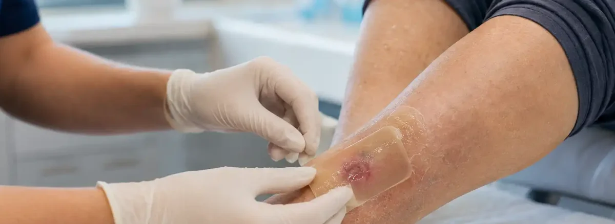
Autolytic debridement is the body clearing dead tissue out of a wound on its own, using enzymes and white cells already present in wound fluid. The dressing over the top cuts nothing and dissolves nothing. It holds moisture against the wound so that process can run.
It is the gentlest method available and the slowest, and it passes through a stage where it looks as though something has gone badly wrong. Yellow sludge appears under the dressing. The wound seems to get bigger. Most people looking this term up are three or four days into a dressing a nurse applied with the instruction to leave it alone. This guide covers what is happening under that dressing, the five signs the process is working, why the appearance deteriorates before it improves, how long clearance usually runs, and the wounds where this method should not be used at all.
On this page
- What is autolytic debridement?
- What the dressing is actually doing
- Five signs it is working
- Why the wound looks worse first
- How long does autolytic debridement take?
- How it compares with the faster methods
- When it is the wrong choice
- The skin around the wound decides a lot
- What sets it back
- Living with a method that takes its time
- When to ask for a reassessment
- Questions people ask
- The takeaway
What is autolytic debridement?
It is the removal of dead tissue from a wound by the body’s own repair machinery, encouraged by a dressing that keeps the wound bed moist. Nothing is cut out, and no chemical is added.
Debridement, by any method, means getting dead tissue out of a wound. Clinical notes often call that tissue necrotic, which means the same thing. That tissue holds healing up in three separate ways: new tissue cannot grow across it, bacteria feed on it, and it hides the real depth of the wound from whoever is assessing it. Clearing it is frequently the step that lets a stalled wound start closing.
The word autolytic comes from roots meaning self-digesting, which describes the mechanism exactly. Wound fluid carries enzymes that break proteins apart, along with white blood cells whose job is to consume debris. Keep that fluid in contact with dead tissue and the tissue is gradually taken apart, over days.
What earns this method its place, despite the slowness, is selectivity. Those enzymes act on tissue that has already died and leave living tissue alone, so healthy tissue cannot be removed by accident, a risk that exists any time a blade is involved. Autolytic debridement is also painless in most cases, needs no anesthetic, calls for no specialist skill at the bedside, and carries on quietly at home between appointments.
Three conditions have to hold. The wound bed needs moisture, the person needs a functioning immune system, and the wound must not be one where infection is spreading. Remove any of the three and the process either stalls or becomes the wrong plan.
What the dressing is actually doing
Far less than people assume. The usual reading is that the gel inside the dressing eats through the dead tissue, the way a drain cleaner works on a blockage. It does no such thing.
The dressing supplies an environment, not an action. Its whole purpose is to keep the wound from drying out, because the enzymes doing the work need water to function at all. A wound bed that has dried into a hard plate is a wound where nothing is happening: no breakdown of dead tissue, and no healing either.
Two families of dressing are used, and the difference between them is the direction moisture travels. Hydrogels add water to a wound that has too little, which makes them the usual pick for a hard, dry, leathery covering. Hydrocolloids, thin films and some foams work the other way, sealing in the fluid a wound is already producing so it stays where it is useful. Both are described as moisture-retentive, and both are ordinary supplies instead of anything exotic.
Matching the dressing to the wound is most of the skill. Put a moisture-adding gel on a wound that is already soaking through and the surrounding skin pays for it; seal a bone-dry crust under a film that adds nothing and a week later it will be exactly as dry. A clinician may also score the surface of a thick dry covering in a crisscross pattern with a scalpel, which sounds dramatic and is simply a way of letting moisture reach the tissue underneath.
This also explains a common reason the method appears to fail. A dressing that dried out, lifted at one corner, or got changed so often the wound never stayed damp for long has not been given the one thing it needed to do its job.
Five signs it is working
Progress here is easy to miss, because every early sign of it looks like deterioration to an untrained eye. Knowing the sequence in advance changes what you see at a dressing change.
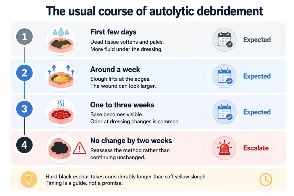
Softening comes first, and it is a change you feel more than see. A covering that was firm and leathery at the start begins to give slightly under a gloved fingertip, and its color usually shifts from dark and matte toward paler and shinier as it takes up water.
Separation follows, and it runs from the outside in. A thin line appears around the rim where dead tissue is parting from living tissue underneath, so the covering starts to look like an object sitting on the wound instead of part of it. The middle stays attached longest, which is why a patch that has clearly lifted at the edges can still be firmly anchored in the center a week later.
Then the volume of fluid climbs. Solid dead tissue is being converted into liquid, and that liquid has to go somewhere, so dressings fill faster and may need changing sooner than they did at the start. Rising drainage in the first week or two of this treatment usually means the method is working, which is the opposite of what rising drainage means in most other situations.
The fourth sign is the one people wait for: red or pink tissue appearing through the covering in small patches, spreading into a continuous surface as more of it clears. A healthy wound base is red or deep pink, slightly bumpy, and moist. Seeing any of it confirms that the layer above was dead tissue and not the wound itself.
The fifth sign reads as bad news and is not: the recorded measurements grow. Dead tissue shrinks and contracts as it dries, pulling the wound edges toward each other and making the whole thing look smaller than it is. Take the covering away and the wound relaxes to its true dimensions. The first measurement taken over a clean base is the first accurate one, and a jump on the chart at that visit reflects better information, not a wound that got worse.
Why the wound looks worse first
Every one of those signs is a change in the direction people associate with trouble. More fluid, a stronger smell, a bigger measurement and a wound bed covered in yellow slime would describe a deteriorating wound in almost any other context.
The yellow material causes most of the alarm, and it is the single most useful thing to understand about this method. As dead tissue breaks down under a moisture-retentive dressing it turns into a soft, stringy, yellow or gray gel that pools in the wound and coats the base. It looks like pus. It is not pus; it is dead tissue partway through being dismantled, which is precisely the result the dressing was applied to produce.
Two features separate them in practice. Broken-down tissue rinses away with normal cleaning and the wound underneath looks unchanged or better; pus keeps re-forming and travels with other signs: heat, swelling, redness reaching out from the edge, pain that is climbing instead of steady. Which of those changes count, and how much weight each one carries, is covered under how to tell if a wound is infected. It is the company the fluid keeps that decides the question, never the fluid on its own.
Odor deserves the same treatment. A dressing sealed against warm skin for several days, with softening tissue inside it, will smell when it comes off, and hydrocolloids have a reputation for a distinctly sour smell of their own. Judge the smell after the wound has been cleaned, not at the moment the dressing is peeled back. An odor that survives cleaning is a finding to report; one that leaves with the old dressing and the rinse is a property of the packaging.
People give up on this treatment during that middle stretch far more often than at any other point, and usually because of how it looks instead of anything a clinician would call a problem. Knowing the sequence in advance is what carries a household through it.
How long does autolytic debridement take?
Days for the first visible change, and weeks for a wound bed that is genuinely clear. This is the slowest method in routine use, and the timeline is the trade for how gentle and how selective it is.
A workable expectation for most wounds: some softening or edge separation inside the first three to five days, meaningful clearance across one to three weeks, and longer where there is a great deal of tissue to get through. In head-to-head research summarized in the National Library of Medicine’s reference chapter on wound debridement, clearing a wound with a hydrogel under this approach took substantially longer than the alternatives it was compared against, a useful corrective for anyone expecting a finished result in a week.
Five factors move the timeline more than the choice of brand. The volume of dead tissue is the obvious one, since a coin-sized patch of soft slough is a different proposition from a thick plaque across a whole heel. Moisture control comes next, because every hour the wound spends dry is an hour of nothing. Blood supply matters, as enzymes and white cells arrive through the circulation and a limb short of flow delivers fewer of both. Immune function matters for the same reason, which is why illness, certain medications and chemotherapy can slow it noticeably. And nutrition sits underneath all of it, since the whole process is work the body funds from what the person eats.
Set a checkpoint at the start instead of waiting to see. Ask at the first dressing change what should have changed by the next appointment, and at what point the plan would be reconsidered. A reasonable rule of thumb is that a week of no visible change at all (no softening, no lifting at the edges, no increase in fluid) is a conversation, not a reason to keep going and hope.
How it compares with the faster methods
Clearing dead tissue can be done in several ways, and they differ mainly in speed, in discomfort, and in how much healthy tissue they put at risk.
Cutting the tissue away with a scalpel or curette is by far the quickest, and it is the route taken when a wound needs to be clean today: a deep infection, an urgent question about how far the damage reaches, or a wound being prepared for surgery. It takes a trained clinician, it can require local anesthetic, and how much comes off at one sitting depends on the setting.
Enzyme ointments occupy the middle ground, applied daily to break dead tissue down chemically from outside instead of relying on the body to supply the enzymes. Sterile medical larvae, used in some centers, are startlingly quick and remarkably selective. Wiping loose material away with gauze or a specialized cloth at a dressing change is the crudest option, and it makes no distinction between what is dead and what is not.
Set against those, this method sits at the slow, patient, unglamorous end of the scale. It asks nothing of the patient beyond time, costs little, and does not attack the tissue that is trying to grow. Those advantages count for most in a person taking blood thinners, someone whose wound is too painful for cutting, a patient receiving comfort-focused care, or a wound being managed at home between clinic visits.
The methods are also not rivals as often as the comparison implies. Softening a hard covering under a hydrogel for several days so it can be cut away comfortably afterward is a routine sequence, and combining approaches is closer to normal practice than picking one and sticking to it.
When it is the wrong choice
Gentle is not the same as universally appropriate. There are wounds where this approach would waste time that matters, and wounds where opening a sealed covering would create a problem that does not currently exist.
An infection that is spreading
Redness traveling out from the wound, swelling, a raised temperature, or a person who has become unwell in themselves changes the priority. Dead tissue is then shelter for bacteria and needs removing quickly, which usually points toward cutting it out. This method may still run alongside other treatment in a wound with local infection, though that is a clinical decision and it comes with closer monitoring.
Dry stable eschar where blood flow is poor
A hard, dry, firmly attached black covering on a heel or a toe, with no redness, softness, fluid or smell around it, is often left exactly where it is. It is acting as a seal over tissue that may not have the blood supply to heal an open wound, and adding moisture to it deliberately converts a closed situation into an open one. The reasoning behind leaving it is set out in our guide to the unstageable pressure ulcer, and what a shortfall in circulation does to any wound on a leg or foot runs through arterial ulcers and ischemia.
Circulation therefore gets checked before dead tissue is moistened on a lower limb. A simple comparison of blood pressure at the ankle and at the arm gives a usable answer in minutes, and how to read that figure is covered in our explanation of the ankle-brachial index.
Too much tissue, or a deadline
A large plaque of thick eschar can take months to shift this way, and months is rarely acceptable when the wound is deep or sitting over bone. Where a skin graft, a flap or a surgical repair is planned, the wound bed has to be clean by a particular date, and waiting for a slow method to arrive there on its own is not a plan.
A weakened immune system
The method depends on the person’s own white cells and enzymes turning up in adequate numbers. Where the immune system is significantly suppressed (by disease, by chemotherapy, by long-term steroids), this approach may simply not deliver, and something that does not rely on the patient’s own biology is often preferred.
The skin around the wound decides a lot
The limiting factor in this treatment is often not the wound itself. It is the border of intact skin around it, which now has to cope with a rising volume of fluid held deliberately in place.
Skin kept wet for days softens, swells and loses strength, a state described as maceration. It shows as a pale white or gray band around the wound edge, wrinkled like a fingertip left too long in the bath, and it may itch or sting. Softened skin tears easily and breaks down under pressure it would otherwise shrug off, so a wound can quietly widen at its margins while the center is doing well. Telling wet damage apart from pressure damage is a skill in its own right, and the two are separated under moisture associated skin damage versus pressure injury.
Three habits protect that border. A barrier film or cream applied to the intact skin, never the wound bed, gives it a waterproof layer to sit behind. The dressing should be sized so the moisture-holding part covers the wound and little else. And a dressing soaking through before its scheduled change is telling you the absorbency no longer matches the amount of fluid being produced, which is a reason to report it instead of adding tape.
Two other things help. Photographs taken at each change, from the same distance and in the same light, turn a vague impression of progress into something a clinician can read. And whatever caused the wound is still causing it. A foot ulcer under this treatment needs the weight kept off it exactly as before, the argument made in full in our guide to offloading a diabetic foot ulcer.
What sets it back
Do not pick at, cut or pull tissue that has started to lift. A partly separated edge is an invitation, and tugging it takes living tissue along with the dead and reopens a surface that was closing. Let the process finish the job, or let a clinician trim what has detached.
Keep antiseptics off the wound unless they were specifically prescribed. Hydrogen peroxide, alcohol, undiluted iodine and bleach solutions damage the same cells this method depends on, so pouring them in works directly against the treatment being given.
Do not open the dressing daily to check. Every change interrupts the moist environment, cools the wound and undoes some of the progress, and the schedule you were given already accounts for how long the dressing needs to stay sealed. Report a dressing that leaks early instead of quietly changing it more often.
Resist the opposite temptation as well. A dressing left well beyond its window because the wound seems to be improving allows fluid to sit against soft skin far longer than intended, and the margins pay for it.
Nothing should be dried out on purpose: no open air, no hairdryer, no leaving it uncovered to form a scab. A dry wound bed halts the process completely. And a hard black covering on a foot or heel should never be softened at home on your own initiative, however tempting it looks, because that decision rests on circulation findings you cannot check yourself.
Above all, do not abandon the treatment on day three because of how it looks. The stage that alarms people is the stage that means it is working, and stopping there returns the wound to where it started.
Living with a method that takes its time
Being told that the plan is to wait is hard to hear, and it is the part of this treatment nobody prepares people for.
Patients and families frequently read a moisture-retentive dressing as an absence of treatment: no procedure, no prescription, nothing to describe when a relative asks what the doctor did. Weeks pass with a wound that looks unpleasant and a plan consisting of leaving it covered. People start to wonder whether they are being managed cheaply, or whether they have simply dropped down someone’s list. That reading is understandable and usually wrong, and asking directly what the dressing is doing and what would make the team change course settles it faster than waiting for reassurance to arrive.
The daily experience has its own weight. Dressing changes bring a smell and an appearance that people find distressing to deal with, and whoever does the changing carries that several times a week. Wounds under this treatment produce more fluid than they did, so clothes and bedding need managing, and social life narrows around the practicalities. Many people stop mentioning any of it, on the grounds that it is temporary and everyone is busy.
Saying it out loud is worth more than it seems. Odor has treatments of its own, including dressings containing activated charcoal. A dressing that fits the household’s routine gets used properly, where one that does not gets skipped. And where the waiting itself has become the problem, a faster method is often available and has simply never been discussed, because nobody realized the slowness was costing anything.
When to ask for a reassessment
Ask for the plan to be looked at again if a week has gone by with no softening, no separation at the edges and no change in the amount of fluid; if the dressing is soaking through well before its scheduled change; if the skin around the wound has turned white, sore or itchy; or if pain has been building steadily from one change to the next.
Some findings need attention the same day: a raised temperature or shaking chills, redness traveling away from the wound, pain climbing quickly, thick discharge with a smell that stays after cleaning, or a dry black covering that has turned soft, wet or boggy. Any of those suggests something happening beneath the surface that the dressing plan was not designed to handle.
One warning sign is easy to attribute to something else. An older person who becomes suddenly confused, drowsy or unlike themselves may be showing the first effect of an infection, and this can appear before any fever does. Where a wound is already being treated, that shift is worth a call the same day.
Questions people ask
Does autolytic debridement hurt?
Usually not. It is generally the most comfortable of the debridement methods, since nothing is cut and no chemical is applied to living tissue. Some people notice mild stinging or itching as the wound bed rehydrates, and removing a dressing that has stuck can be briefly uncomfortable. Pain that is increasing from one dressing change to the next is a different matter and should be reported, because it points more often to infection than to the treatment itself.
Is the yellow material in my wound pus or dead tissue?
Under a moisture-retentive dressing it is most often dead tissue partway through breaking down, which forms a soft yellow or gray gel closely resembling pus. Cleaning tells you a good deal: broken-down tissue rinses off and leaves a wound that looks the same or better, while pus re-accumulates and arrives with heat, swelling, spreading redness or rising pain. Where the two cannot be told apart at home, have the wound seen instead of guessing.
Can autolytic debridement be done at home?
It is frequently continued at home, which is one of its practical advantages, but the decision to use it belongs with a clinician who has assessed the wound. That assessment covers whether infection is present, whether the blood supply can support an open wound, and which dressing suits the amount of fluid involved. Applying a moisture-retentive dressing to a wound nobody has evaluated can do real harm, particularly on a foot.
What is the difference between autolytic and enzymatic debridement?
The source of the enzymes. In the autolytic approach they come from the patient, carried in wound fluid that a dressing keeps in place. In the enzymatic approach a manufactured enzyme is applied to the wound as an ointment, usually daily, and works on dead tissue from outside. The ointment tends to be quicker and needs a prescription; the dressing route is gentler, cheaper and slower. Neither excludes the other, and both are sometimes used alongside a clinician trimming tissue that has loosened.
Does it work on hard black eschar?
It can, with a hydrogel supplying the water that dry tissue lacks, and the covering may be scored first so moisture gets underneath. Whether it should be attempted at all is the more important question. Hard dry eschar on a heel or toe with a poor blood supply is often preserved deliberately instead of softened, because the tissue beneath may not be able to heal an open wound. That judgment follows an assessment of circulation and is not one to make at home.
What happens if it does not work?
The plan changes, and that is a normal outcome instead of a failure. A wound showing nothing after a week or two may be too dry, may be short of blood supply, may carry more dead tissue than this method can handle, or may need an immune system doing better than it currently is. Depending on which applies, the next step might be a different dressing, an enzyme ointment, a vascular opinion, or clearing the tissue with an instrument.
The takeaway
Autolytic debridement asks the body to clear its own dead tissue, and asks the dressing only to keep the wound moist enough for that to happen. It is painless in most cases, it leaves living tissue alone, and it is slower than every alternative.
Expect softening and lifting at the edges within the first few days, more fluid than before, a yellow gel that looks like pus and is not, and red tissue appearing in patches as the covering clears. Expect the measurements to grow at the visit where the base finally becomes visible. Protect the skin around the wound while all of that is going on, because that border is where this treatment usually runs into trouble.
And hold the method to a timetable. A week with nothing to show for it is worth raising, and a wound that is infected, deep, sitting on a poorly supplied foot, or heading for surgery generally needs something faster than patience.
Not sure whether the dressing is working?
A wound that has not changed in a week, or one where the surrounding skin is starting to suffer, is worth having looked at. An assessment can tell whether to carry on, switch dressings, or clear the tissue a faster way.
Choosing between the methods, and matching a dressing to how much fluid a wound is producing, is part of our advanced wound care service, where the reason the dead tissue is there in the first place gets examined alongside the plan for removing it.
This article is general education and does not replace a clinical assessment. The debridement method that suits a wound depends on infection, blood supply and the amount of dead tissue present, and no moisture-retentive dressing should be applied to a wound that has not been evaluated by a healthcare professional.