Moisture associated skin damage is skin worn down by staying wet, and it is the thing most often mistaken for a bedsore. Both turn up on the same people, in roughly the same territory, and both announce themselves as a patch of discolored skin. They come from opposite directions, and almost nothing that fixes one does anything for the other.
Sorting them out is not a matter of vocabulary. A pressure injury needs the pressure taken off. Moisture damage needs the skin kept dry and shielded. Guess wrong and the wound sits there while somebody diligently applies the wrong remedy. This guide covers what the damage actually is, why the mix-up happens so reliably, the three features that separate the two, the four places moisture damage turns up, and the point where either one stops being something to manage at home.
On this page
- What is moisture associated skin damage?
- Why the two get read as each other
- Three signs that separate them
- The four places it shows up
- Why the press test is not the whole answer
- When either one needs attention
- What actually helps
- What to avoid
- Living with it
- When to have it looked at
- Common questions
- The takeaway
What is moisture associated skin damage?
Moisture associated skin damage is inflammation and erosion of the skin caused by long contact with fluid: urine, stool, sweat, wound drainage, or output leaking from a stoma. Clinicians shorten it to MASD, and you may also see it written with a hyphen as moisture-associated skin damage.
The outer layer of skin is a barrier engineered to work dry. Leave it soaking and three things happen at once. It swells and softens, a change called maceration, which makes it weak enough to tear under a load it would normally shrug off. Urine pushes its acidity in the wrong direction, and the enzymes in stool go to work on the fats that hold the surface cells together. Skin in that state does not need much to break. A bedsheet dragging across it during a transfer is plenty.
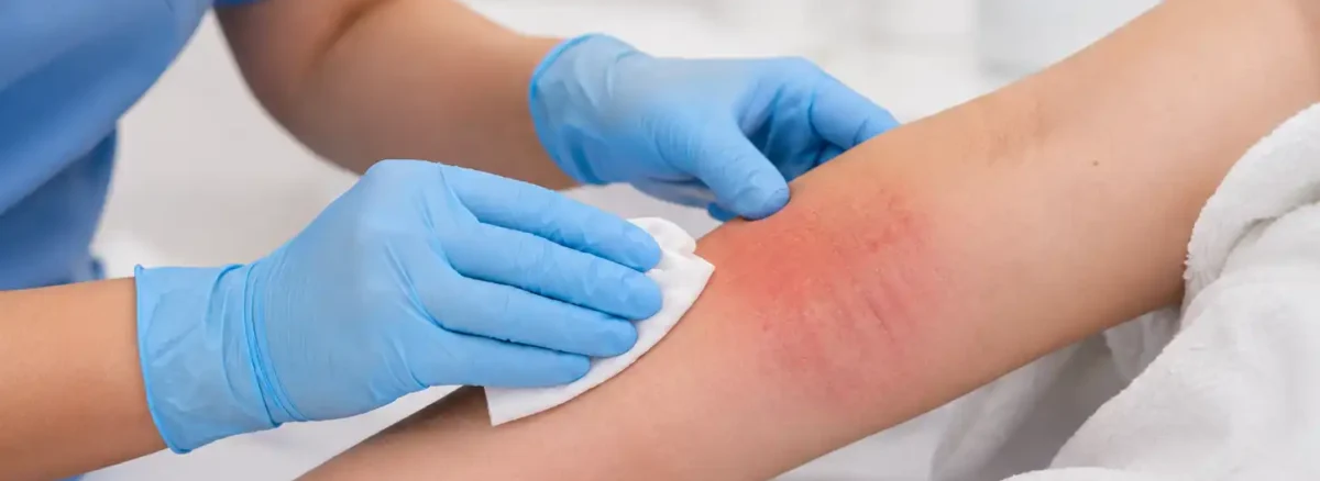
What you see is a patch that looks red on pale skin, and darker, duller or purplish on brown and black skin. It often looks wet or shiny even after drying. Sometimes the surface goes white and waterlogged. Where the top layer has already rubbed away, there is a shallow raw area that weeps. People describe it as burning or stinging, and it usually stings hardest during cleaning.
Hold on to one fact and much of the confusion resolves itself: this is a surface injury. It starts at the top of the skin and works inward.
Why the two get read as each other
A pressure injury travels the opposite way. Sustained pressure squeezes tissue between a bone and a mattress or chair, blood supply falls, and cells starve. Much of that harm can begin deep, near the bone, and take days to surface. By the time it is visible, the damage underneath may be older and larger than the mark on top suggests.
Outside-in against inside-out, and yet the two arrive looking almost identical, because everything around them is shared. They happen to the same people: someone immobile, someone incontinent, someone whose skin is thin with age or illness. They occupy the same real estate, since the buttocks and the base of the spine are simultaneously the bonier region of a seated body and the wettest. And on day one, both are simply an area of skin that has changed color.
They also encourage each other. Damp skin has a higher friction coefficient than dry skin, meaning it grips and drags instead of sliding, so shear forces bite harder during every reposition. Softened skin also tolerates less pressure before it gives. Moisture is significant enough on this count that it is scored as its own item on the risk assessment tools nursing teams use at admission.
Finding moisture damage does not rule out a pressure injury. Both can sit on the same square of skin, and in bedbound patients that combination is ordinary. The question worth asking is rarely which one it is, but how much of each is in play.
Three signs that separate them
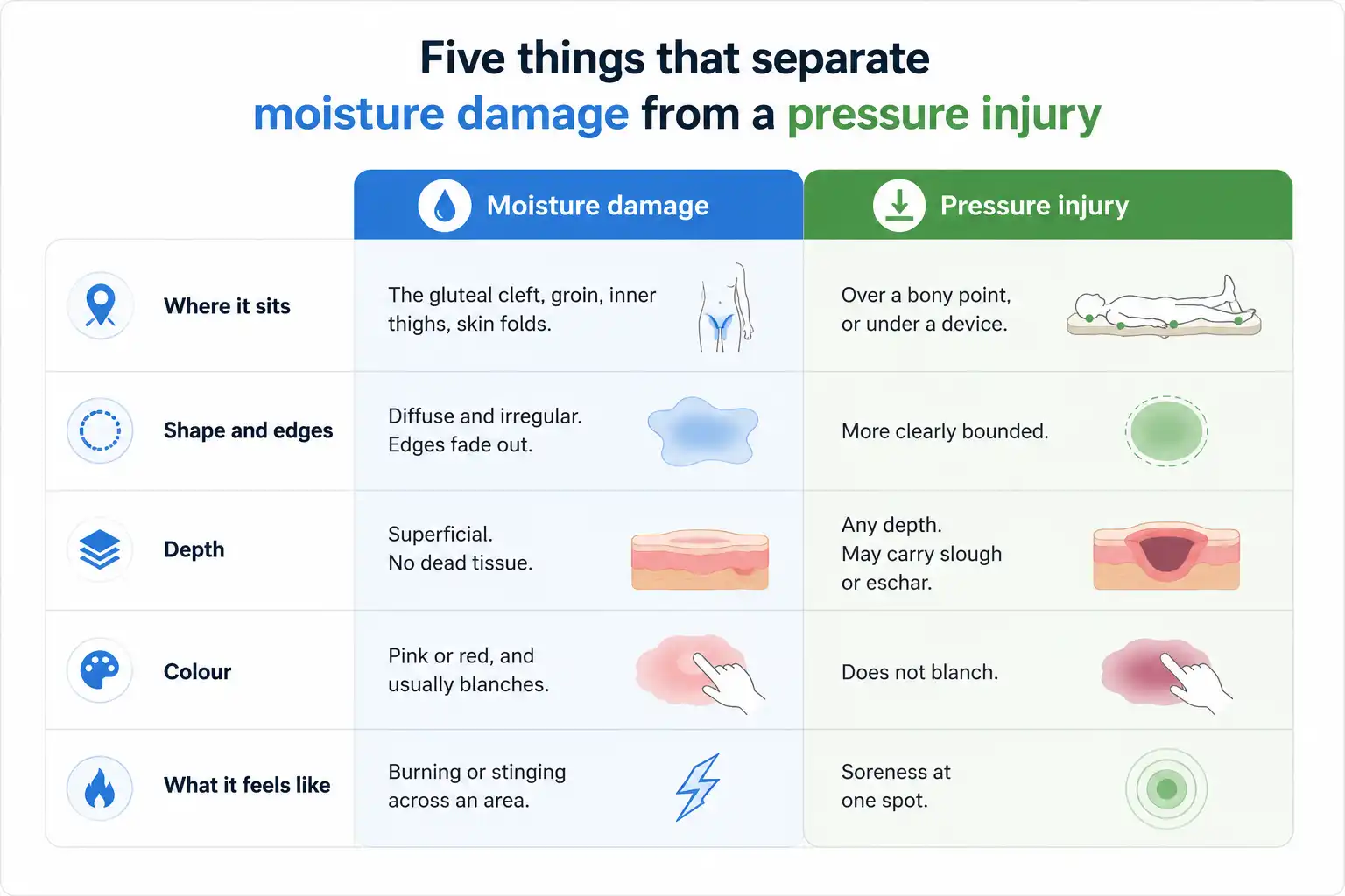
Where it sits, what its edges look like, and how deep it goes. Those three read the difference more reliably than anything else available without equipment.
Where it sits
Pressure injuries form where bone presses skin against something solid: the tailbone, the heels, the hips, the sitting bones, the elbows, the back of the head. They also form under equipment: oxygen tubing behind the ears, a catheter lying against a thigh, the rim of a cast or a brace.
Moisture damage forms where fluid pools and cannot evaporate: the crease between the buttocks, the groin, the inner thighs, beneath the breasts, under a fold of abdominal skin, in the creases of the neck, around a stoma, and on the ring of skin surrounding a wound that drains heavily.
The lists overlap at exactly one awkward place, the sacrum and buttocks, which is where most arguments about this happen. Location narrows the field. The edges finish the job.
What the edges look like
A pressure injury tends to be one patch with a findable outline, roughly reproducing the shape of whatever was pressing on it. Trace it with a finger and you can say where it stops.
Moisture damage sprawls. Its borders are vague and blotchy and dissolve into normal skin without a clear frontier. It often appears as several patches at once, and it is frequently mirrored on both sides of a fold, because the two facing surfaces sit in the same wetness and take the same insult. Nurses call those kissing lesions, and a matched pair either side of a crease is close to a giveaway.
One extra clue lives at the perimeter. Small separate spots scattered a short distance beyond the main red area usually mean yeast has settled into the damaged skin, which changes what the area needs.
How deep it goes
This is the decisive one. Moisture damage stays shallow. It strips the upper layers and can leave a raw, weeping, tender area, but on its own it does not carve a crater, expose fat or muscle, or manufacture dead tissue.
So a wound with visible depth, a rim of yellow stringy material, or a dry brown or black plaque across it was not produced by wetness alone. Those are features of deeper injury, and the four-level system used to describe them is laid out in pressure ulcer stages. Where dead tissue covers the base completely, nobody can measure the depth until it is cleared, a situation described under unstageable pressure ulcer.
The four places it shows up
MASD is an umbrella covering four situations. They share a mechanism and differ in which fluid is doing the harm, which decides what has to change.
Incontinence-associated dermatitis comes from urine, stool, or both, and it is far and away the most common version. It covers the perineum, the buttocks, the inner thighs and the groin. Stool is the more aggressive of the two, since digestive enzymes carried in it keep working on skin after they have finished with food, and liquid stool spreads further and clings longer than formed stool.
Intertriginous dermatitis is sweat sealed inside a skin fold with nowhere to evaporate. Under the breasts, in the groin, beneath an abdominal apron, in the folds of the neck. Heat and friction build alongside the wetness, the mirrored kissing pattern is at its most obvious here, and yeast joins in often enough that a persistent fold rash is treated as fungal until shown otherwise.
Periwound maceration is wound fluid attacking the healthy skin around a wound because the dressing is not absorbing everything the wound produces. That skin turns white and boggy, the wound edge softens, and the wound quietly gets wider while the middle looks fine. Heavily draining leg wounds are the usual setting, and the interaction between drainage, dressings and compression bandaging is covered in our page on the venous leg ulcer.
Peristomal moisture damage is stoma output creeping under the appliance and lying against skin. The skin treatment matters, but the fix is the seal. A pouch that keeps leaking needs refitting, and no amount of cream compensates for an appliance that does not sit flat.
Why the press test is not the whole answer
The check most people are taught is simple. Press a finger gently on the discolored skin for a few seconds and lift it. If the spot pales and then refills, the redness blanches. If it stays the same color under the finger, it does not.
Redness that refuses to blanch, sitting over a bony point, is the textbook definition of the earliest stage of pressure injury. Moisture-related redness more often blanches. As a rule of thumb it earns its keep on pale skin.
On brown and black skin the test loses much of its value, because redness is not the change to look for in the first place. Early pressure damage there may read as an area darker than its surroundings, or purple, or simply flatter and duller in tone.
Better signals come from elsewhere. Compare the area with the skin immediately beside it in good daylight. Lay the back of a hand on it, then on the matching spot on the other side, and feel for a temperature difference. Press for firmness, or for a soft boggy give underneath. And ask about pain, itching or tingling, since discomfort commonly arrives before anything is visible at all.
Skipping those steps is one reason early pressure damage can go unrecognized for longer on darker skin and be found at a more advanced point. A caregiver who checks by touch and by comparison, not by looking for red, catches considerably more.
When either one needs attention
Most moisture damage settles within days once the skin stops being wet. What follows is the short list of things that mean the situation has moved past routine skin care.
An area that keeps enlarging after several days of consistent barrier protection is telling you something is still soaking it, or that the diagnosis needs revisiting. Scattered spots creeping outward from the main patch point at yeast, which needs an antifungal and gets no benefit from more barrier cream. Skin that has broken open and weeps steadily has stopped being irritation and become a wound.
Infection changes the picture again. Warmth spreading past the damaged skin, swelling, pain that climbs instead of easing, thick or foul drainage that persists after the area is cleaned, or a raised temperature all point beyond the skin surface, and the fuller set of signals is gathered in how to tell if a wound is infected.
Anything purple or maroon, a blister filled with blood, a firm boggy patch, a dry black or brown plaque, or a crater with yellow material in it belongs to a different category entirely. Wetness does not create those. They should be looked at by a clinician, and they should not be treated as a rash that will pass.
A fever or shaking chills, redness traveling outward quickly, pain rising fast, or someone suddenly seeming unwell in themselves needs same-day medical attention.
What actually helps
Work in order, because the steps are not interchangeable. Source first, then cleaning, then barrier, and keep the pressure plan running underneath all three.
Dealing with the source is the step people skip, and it decides everything downstream. That means an absorbent product designed to pull fluid away from skin instead of holding it against the body, changed as soon as it is soiled and not at the next scheduled round. It means treating the reason for diarrhea where there is one. It means a stoma appliance that seals, and a dressing matched to how much a wound is producing when drainage is the culprit. Skin cannot repair while it is still being soaked.
Cleaning should be gentle and infrequent enough to leave the skin alone between times. A no-rinse cleanser formulated for skin, or one balanced to skin’s natural acidity, treats the surface better than repeated washing with ordinary soap, which is alkaline and strips oils that are already depleted. Pat dry. Never rub. Let a fold open to the air for a few minutes before closing it.
Then the barrier, in a thin layer. Zinc oxide, dimethicone, petrolatum ointments and liquid barrier films all work by keeping fluid off the skin. Thickness is where this goes wrong: a heavy coat blocks the absorbent product from doing its job and can leave the area wetter, so apply enough to see a sheen and no more. Reapply according to the product’s instructions instead of after every single change, unless the label says otherwise.
Underneath all of it, the pressure plan carries on regardless. Repositioning on a schedule, floating heels clear of the bed, and using a proper support surface are unrelated to skin barrier products and cannot be substituted by them. MedlinePlus, the patient-facing service run by the US National Library of Medicine, sets clean dry skin and repositioning every two hours next to each other in its guidance on pressure sores, and that pairing is the one to copy at home.
What to avoid
Keep powder out of skin folds. Talcum powder and cornstarch both cake into gritty clumps once they meet sweat, and those clumps then abrade the very skin they were meant to protect. Cornstarch has a second problem, in that yeast will happily grow on it.
Do not scrub, and do not rub dry with a towel. Waterlogged skin tears at a fraction of the force normal skin takes, and friction supplies the second half of the injury that wetness started. The same logic rules out wipes containing alcohol or fragrance on skin that is already broken, which sting badly and slow repair.
Resist the urge to layer protection. An incontinence pad on top of a draw sheet on top of a plastic undersheet traps heat and humidity against the body and stops a pressure-redistributing mattress from contouring to it, so two products meant to help cancel each other out. One correctly chosen absorbent product beats three stacked ones.
Do not leave a soiled pad in place until the next scheduled check, and do not reduce someone’s drinking to cut down on urine. Dehydration makes skin more fragile, concentrates urine so it irritates more, and works against healing everywhere else in the body.
Finally, do not let the argument over which injury it is hold up the response. Controlling moisture and relieving pressure are both harmless to someone who turns out not to need one of them. Do both while you wait for a clinician to settle it.
Living with it
Almost nobody writes about the part families actually struggle with, which is the blame.
A relative caring for someone at home discovers a red patch, hears the word bedsore, and concludes they caused it through neglect. Sometimes a visiting professional implies as much. It is worth saying plainly that skin damage from incontinence can appear within hours on fragile skin, especially after an episode of diarrhea overnight, and it appears on people whose care is careful and constant. Skin that has become thin with age, illness, steroids or poor nutrition gives way to insults that would not have marked it a decade earlier. Discovering damage is a reason to change the plan, not evidence of a failure.
For the person it is happening to, the dominant experience is stinging, and it peaks precisely when someone is cleaning the area. That sets up a loop worth breaking early: cleaning hurts, so it gets rushed or postponed, so the skin stays wet longer and hurts more the next time. Say clearly if cleaning is painful. Barrier films that go on without friction, gentler cleansers and timing pain relief before care are all things a nurse can arrange.
Embarrassment does its own damage. Incontinence is the part people conceal hardest, which means the skin problem it causes gets described vaguely or not at all, and some people cut back on fluids in a private attempt to manage it. Neither helps. If it is easier to write it down than say it out loud, write it down and hand it over.
Two practical habits are worth building. Look at the skin properly in daylight during one care episode a day, comparing left with right, and photograph anything that changes so a clinician can see the progression instead of a single snapshot. Then ask outright whether a barrier product and a repositioning schedule are both on the care plan, since it is common for one to be there and the other to be assumed.
When to have it looked at
Arrange an appointment if a red or discolored patch has not started improving after two or three days of consistent moisture control and barrier protection, if the skin has opened at any depth, if spots are spreading outward from the edges, or if pain seems out of proportion to what is visible.
Anything purple, maroon, black or brown, and any blister, deserves assessment instead of watchful waiting, because those appearances belong to deeper injury and the surface almost always understates them.
Seek same-day care for fever or chills, redness moving outward quickly, pain climbing hour by hour, or a sudden general decline in how someone seems. Those suggest infection that has spread past the skin.
One further rule covers the uncertain cases. If the person spends most of the day in a bed or chair and the affected skin lies over a bone, assume a pressure injury may be involved and act on that assumption until someone qualified rules it out. Skin checks and dressing changes for people being cared for at home fall within our mobile and home health wound care service.
Common questions
Is moisture associated skin damage the same as a bedsore?
No. A bedsore, properly called a pressure injury, is caused by sustained pressure cutting off blood supply between a bone and a surface. Moisture damage is caused by fluid sitting on skin and breaking down its outer barrier. They look similar early on and often occur together, but they need different treatment.
Can moisture damage turn into a pressure ulcer?
Not by itself, since the two have different causes. Wet, softened skin is more vulnerable to pressure and shear, so an area with moisture damage can develop a pressure injury more readily than intact skin would. Controlling the moisture is therefore part of preventing pressure injuries, not a separate task.
What does moisture associated skin damage look like?
Typically a poorly outlined patch of discoloration with a wet or shiny surface, sometimes white and waterlogged, sometimes raw where the top layer has gone. It frequently appears on both sides of a skin fold at once. Where skin tone is deeper, the change may register as darkening or dullness with no obvious redness at all.
How long does it take to heal?
Uncomplicated cases often improve within a few days and settle over one to two weeks once the skin is kept dry and protected, though this varies with the person’s general health, nutrition and how completely the moisture has been controlled. Skin that shows no improvement after several days of consistent care should be reassessed.
Should I use barrier cream and powder together?
No. Powder mixed with cream forms a gritty paste that rubs skin and hides the area from view. Choose one barrier product, apply it thinly, and follow the instructions on it. If a fold keeps sweating, a moisture-wicking textile designed for that purpose is a better answer than powder.
Can it happen to someone who is not incontinent?
Yes. Sweat trapped in a skin fold, drainage from a wound soaking the skin around it, and leakage under a stoma appliance all produce the same damage. Continence is only one of the four routes, and a fold rash under the breasts or abdomen is a common presentation in people with no continence problem at all.
The takeaway
Moisture associated skin damage is a surface injury caused by wetness, and it is not a bedsore. Check three things and you will usually know which you are dealing with: whether the skin sits in a fold or over a bone, whether the edges are blurred or definable, and whether there is any real depth to it. Depth settles it, because wetness alone cannot dig.
Treatment follows the cause. Stop the skin getting wet, clean it gently, protect it with a thin barrier, and keep repositioning on schedule whichever injury it turns out to be. What should prompt a call is depth, darkening color, spreading spots, or fever alongside any of it.
Not sure whether it is moisture or pressure?
An assessment can tell the two apart, check whether anything deeper is developing underneath, and set out a skin plan that covers both causes at once.
Skin that keeps breaking down in the same place, whatever the cause, is handled through our chronic wound treatment service, where the assessment covers the moisture, the pressure and the reasons the skin is failing to hold.
This article is general education and does not replace a clinical assessment. Discolored, broken or painful skin in someone with limited mobility should be examined by a healthcare professional, particularly where the area has depth, is changing color, or appears alongside a raised temperature.