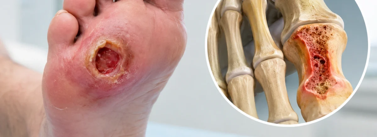
An osteomyelitis foot ulcer is a foot wound where infection has traveled out of the soft tissue and into the bone beneath it. The signs that this has happened are far quieter than most people expect. Often there is no fever, no dramatic change on the surface, and no extra pain at all.
That silence is what makes bone infection so easy to miss in a foot. The wound looks much as it did last month, the person feels much as they did last month, and the thing keeping the ulcer open sits a few millimeters below the wound bed. This guide covers what bone involvement means for treatment, the six signs that raise suspicion, the bedside check clinicians reach for first, why an early X-ray can reassure everybody wrongly, and how the diagnosis is finally settled.
On this page
- What an osteomyelitis foot ulcer is
- Why the surface of the wound tells you so little
- Six signs a wound may have reached the bone
- The bedside check clinicians reach for first
- Why an early X-ray can be falsely reassuring
- How the diagnosis is confirmed
- Treatment, and why the antibiotic course varies
- What you can do while this is being sorted out
- Mistakes to avoid
- Living with a bone infection diagnosis
- When to be seen urgently
- Common questions
- The takeaway
What an osteomyelitis foot ulcer is
Osteomyelitis means infection inside a bone. In the foot it almost always arrives the short way: bacteria in an open wound work downward through the soft tissue until they reach the bone directly below. Elsewhere in the body bone infection usually travels through the bloodstream. In a foot ulcer the route is a few millimeters of tissue, which is why depth is such a strong clue.
The distinction changes everything about treatment. Soft tissue infection responds to a normal course of antibiotics because blood carries the drug easily into inflamed flesh. Bone is denser, less well supplied, and can hold pockets of dead material that no antibiotic reaches at a useful concentration. Bacteria also settle into a protective layer on the bone surface that shields them from the immune system and the medication alike.
The practical consequence is simple and easily missed: an ulcer sitting on infected bone will not close, however good the dressing regimen is. Weeks of careful wound care pass with steady, unremarkable non-progress. The dressings were doing their job. The problem was one layer down.
Why the surface of the wound tells you so little
Most people assume that an infection in the bone would announce itself. It would hurt more, the foot would look angrier, and there would be a temperature. All three assumptions fail regularly in the foot, and each fails for a different reason.
Pain fails first. Long-standing diabetes commonly damages the nerves supplying the feet, and damaged nerves report badly. Somebody whose foot has lost sensation can walk around on an infected bone without any signal that something has changed. The alarm has been disconnected, and its silence gets read as an all-clear.
Temperature fails next. A raised temperature and a high white cell count are what people expect from a serious infection, and plenty of people with bone involvement in a foot ulcer have neither. The infection is walled off in a small volume of bone and the rest of the body stays largely unbothered by it.
Appearance fails last, and it fails in a specific way. The opening you can see is the narrow end of the problem. A wound the width of a fingertip can sit above a tract running down to bone, because skin at the surface closes in from the edges while the deeper channel stays open. A small ulcer is not a shallow one, and the two get confused constantly.
So the useful question is not how bad the wound looks. It is how deep it goes, how long it has been there, and what sits underneath it.
Six signs a wound may have reached the bone
None of these confirms anything alone. Together they are what makes a clinician stop treating a wound as a skin problem and start investigating the bone.
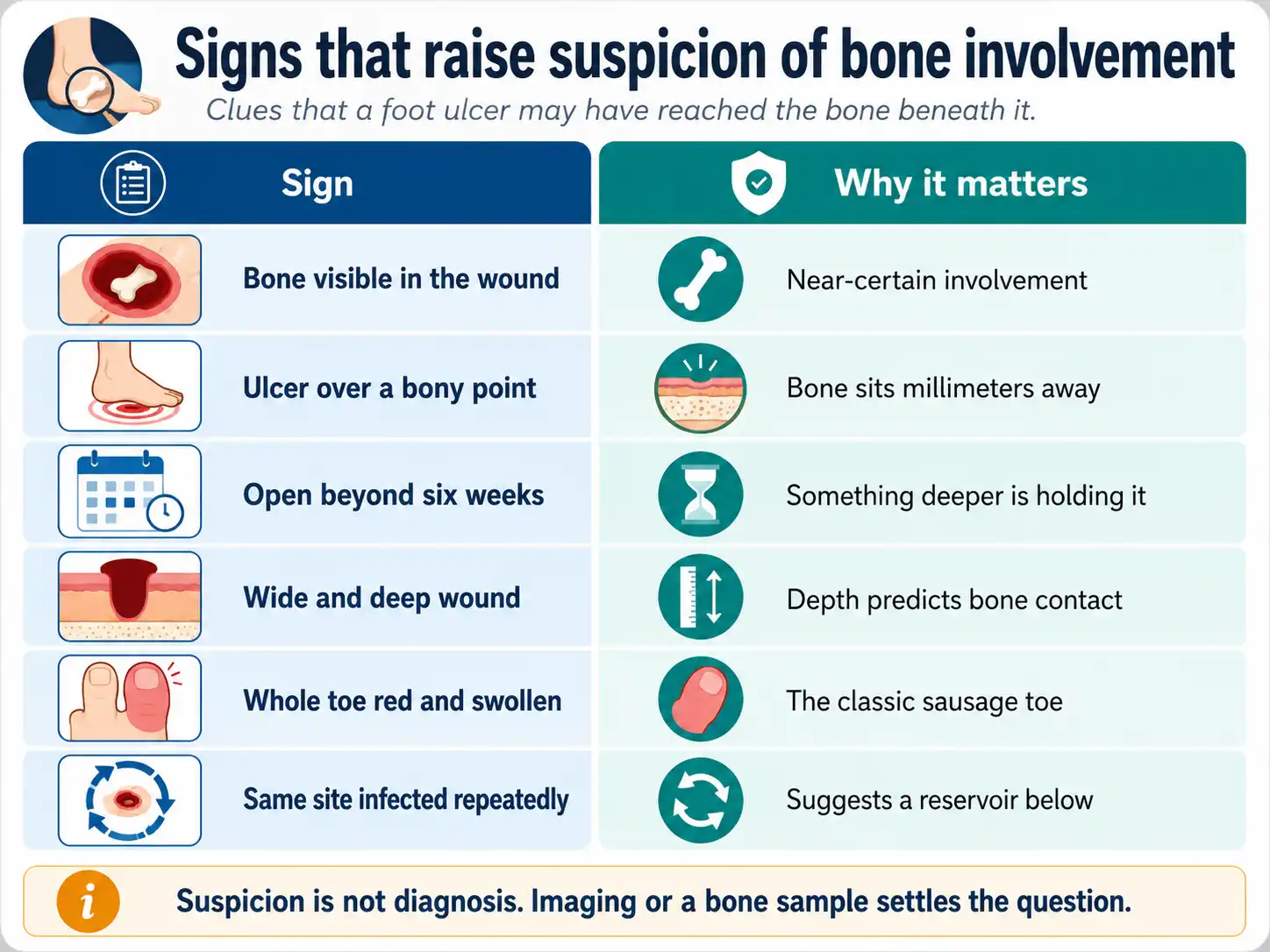
Visible bone in the wound base is the least ambiguous of the six. Bone exposed to open air in a chronically infected wound is assumed to be involved until proven otherwise, and it moves the conversation straight to imaging.
Position carries more weight than most people realize. An ulcer over the ball of the foot, the tip or knuckle of a toe, the heel or the outer edge has very little tissue between it and bone, while the same wound on the fleshy arch has centimeters of protection. Where a wound sits changes how alarming its depth is.
Duration is the sign people underestimate most. A foot wound still open beyond about six weeks despite genuine treatment is signaling that something is obstructing it, and bone infection sits high on the list of candidates. Size and depth work the same way: figures often quoted in diagnostic guidance put ulcers larger than around two square centimeters, or deeper than around three millimeters, into a higher-risk group.
The sausage toe has an unattractive name and a useful meaning. One toe uniformly red, swollen and shiny along its whole length, with normal toes on either side of it, is a recognized presentation of bone infection in that toe.
Repetition finishes the list. A wound that heals, breaks down at the same spot, takes a course of antibiotics, settles and breaks down again is behaving like a wound with an untreated source beneath it. Each episode looks manageable on its own. The pattern across a year does not.
The bedside check clinicians reach for first
Before any scan, there is a check that takes about fifteen seconds and costs nothing. It is called the probe-to-bone test.
A clinician passes a sterile blunt-tipped metal probe gently into the wound, following whatever channel is already there without forcing a new one. If it meets a hard, gritty surface at the bottom, that surface is bone. If it meets soft resistance and stops, it has not.
How much the result means depends on who is being tested. In somebody with a long-standing diabetic foot wound, where bone infection was already plausible, touching bone raises the likelihood substantially and usually triggers imaging. In a fresh, shallow wound on a foot with good sensation and good circulation, not touching bone is genuinely reassuring.
This is not something to attempt at home. Pushing anything into a wound risks driving surface bacteria deeper, tearing the tissue trying to bridge the gap, and creating a false channel that was never there. Ask whether the test has been done; leave the doing of it to somebody trained.
Why an early X-ray can be falsely reassuring
Here is the part patients are almost never told, and it explains a sequence of events that feels like a mistake but usually is not one.
A plain X-ray is normally the first image taken, because it is cheap, fast and available everywhere. What it shows is bone density. An infection has to destroy a substantial share of the mineral in a bone before the loss becomes visible on film, and that takes time, commonly a couple of weeks or more from the moment the infection took hold.
So an X-ray taken early can look entirely normal while the bone is already infected. Everybody relaxes, the wound gets another six weeks of dressings, and a repeat film later shows changes that were building the whole time. Nothing was overlooked; the imaging was asked a question it could not yet answer.
Repeating a plain X-ray two to four weeks after a normal one is a recognized approach for this reason, and the second film is frequently the informative one. If a first X-ray comes back clear on a wound that is not healing, the question to ask is when the image will be repeated, or whether it is time for a scan that does not depend on mineral loss.
How the diagnosis is confirmed
Three tools do most of the work, and each answers a different question.
MRI
An MRI scan looks at the marrow inside the bone instead of the mineral around it, so it picks up infection considerably earlier than plain film. It also maps how far the problem extends, which is what a surgeon needs before deciding how much bone has to go. Its limitation is that inflammation from other causes looks similar, and in a foot also damaged by the bone and joint collapse that diabetes sometimes causes, telling the two apart takes experience and often more than one test.
Bone biopsy and culture
Taking a small sample of the bone itself and growing it in the laboratory is the reference standard. It answers what imaging cannot: whether the bone is genuinely infected, and precisely which organism is responsible along with the antibiotics it responds to.
That second answer is worth more than it sounds. A swab taken from the surface of an ulcer grows whatever is living on the surface of an ulcer, which is usually a mixed population of bacteria that may have nothing to do with what is in the bone. Treating the bone based on a surface swab is a well-recognized way to end up on the wrong antibiotic for six weeks. If a wound is not responding as expected, asking whether the bone itself has been sampled is a reasonable and specific question.
Blood tests
Markers of inflammation in the blood support the picture without settling it. A markedly raised marker in somebody with a deep, chronic foot wound adds weight to the suspicion, and normal results do not clear the bone. They are most useful watched over time, since a marker falling steadily through treatment suggests the treatment is working.
Treatment, and why the antibiotic course varies
Two broad routes exist, and the choice between them shapes everything that follows, including how long the antibiotics run for.
The first is antibiotics alone, chosen where the infected bone is limited, blood supply to the foot is adequate and surgery carries more cost than benefit. Because the drug has to penetrate bone and finish the job unaided, these courses are long. Six weeks is a common figure, sometimes starting intravenously and switching to tablets once things settle.
The second route removes the infected bone surgically, and the antibiotic course afterward is judged by what is left behind. When a surgeon is confident all the infected bone has been taken out, the remaining tissue is being treated as a soft tissue infection and a short course, sometimes little more than a week, may be sufficient. When infected bone had to be left in place, the long course returns. The single question that most influences the length of your antibiotic course is whether infected bone was removed.
Surgery here spans a wide range: shaving a small area of bone through a wound that is already open, removing a single bone within a toe, or removing the toe. That last option lands hard when it is first raised, and the reasoning behind it is worth hearing. A toe removed cleanly can give a foot that heals in weeks and stays stable for years, where the alternative is sometimes months of treatment and a wound that reopens. The decision is made with the whole foot in mind.
Blood supply runs underneath both routes. An antibiotic only works where blood delivers it, so circulation to the foot is usually assessed early, and improving it can be the step that makes everything else effective. Once the infection is controlled the wound above it still has to close, and pressure relief decides that part; how weight is taken off a healing wound is set out in offloading a diabetic foot ulcer.
What you can do while this is being sorted out
More than it feels like, though most of it is undramatic.
Keep weight off the wound every time, including the short trips around the house that feel too brief to count. Take the antibiotic course to the last dose. Keep blood glucose as steady as your team advises, since high glucose blunts the immune cells doing the fighting. If you smoke, stopping produces a measurable return here, because nicotine narrows the small vessels carrying oxygen and antibiotic into the foot.
Keep a simple record. A weekly photograph from the same distance, plus a note of any change in drainage, smell, swelling or how the foot feels, gives your team a timeline that memory cannot reconstruct in an appointment.
And ask three specific questions, because the answers shape everything and are rarely volunteered. Which bone is affected? Was a sample of it taken, or is the antibiotic based on a surface swab? What is the plan if this course finishes and the wound has not closed? People who know those answers cope far better with a long treatment than people waiting to be told.
Mistakes to avoid
Do not probe, poke or explore the wound yourself to find out whether you can feel bone. The test is safe in trained hands because of how it is done, and unsafe otherwise for exactly the same reason.
Do not stop antibiotics early on the strength of an improved-looking wound. Skin closing over infected bone is a familiar pattern and a deceptive one, and a course abandoned halfway tends to buy a few comfortable weeks followed by a recurrence that is harder to treat.
Do not treat a clear X-ray as the end of the matter when the wound is not healing, and do not treat a surface swab as the identity of the organism in the bone. Both are reasonable starting points and neither is a conclusion.
Do not keep walking on the foot because it does not hurt. Absent pain in a foot with nerve damage is a broken instrument reading, not a reassuring measurement. And do not put off saying that something has changed, since delay is the one factor here that patients control directly and the one most strongly linked to a worse outcome.
Living with a bone infection diagnosis
The clinical plan gets explained carefully. What it will be like to live through generally does not, and that gap is where most of the distress collects.
Treatment is long by the standards of anything else people are used to. Six weeks of antibiotics may involve a fine tube threaded into a vein in the arm so doses can be given at home, regular blood tests, and a run of appointments that eats into work and income. Driving may be restricted. Ordinary shoes may not fit over the dressing. Plans made months ago quietly fall away.
Underneath the practical disruption sits the fear almost nobody says out loud, which is the fear of losing part of the foot. It deserves a direct answer instead of a reassuring silence, and the honest one is specific to your foot: how much bone is involved, and how well blood reaches it. Asking your team outright is the fastest route to that answer, and a real answer is nearly always easier to carry than an imagined one.
The waiting has its own weight. Weeks pass with no visible change, because bone heals slowly and offers no weekly progress report. Ask what a good result would look like at four weeks and at eight, so there is something concrete to measure against instead of a wound that seems to be doing nothing.
When to be seen urgently
Get same-day medical attention for a raised temperature or shaking chills, redness traveling upward from the wound, a sudden increase in swelling or discomfort, drainage that turns foul or much heavier, any tissue turning black, or blood glucose that becomes unexpectedly hard to control. That last one catches people out; a sharp loss of glucose control with no change in diet or medication can be the first outward sign that an infection is escalating.
Arrange a review promptly, though less urgently, for a foot wound still open after about six weeks, one that breaks down repeatedly at the same site, a toe swollen and red along its length, or a wound where you or anybody dressing it can see or feel bone. The National Library of Medicine’s plain-language overview of bone infections is a reasonable place to read further while you wait to be seen.
If you are already being treated for bone infection, report a wound that starts enlarging again, drainage that returns after settling, or a marked change in smell. Recurrence is easier to deal with early, and a mid-course change should not wait for the next appointment.
Common questions
Can bone infection in a foot ulcer heal without surgery?
Often, yes. Antibiotics alone resolve a good number of these infections, particularly where the affected area of bone is small, blood supply to the foot is reasonable and treatment starts before the infection is extensive. Which route suits a particular foot depends on how much bone is involved and how well blood reaches it, which is what the imaging is for.
How long does treatment usually take?
Around six weeks of antibiotics is common where infected bone stays in place. Where all of the infected bone is surgically removed, a much shorter course may be enough. The wound above the bone frequently takes longer than the antibiotic course to close, so treatment finishing and the ulcer healing are two separate milestones.
Does bone infection mean I will lose my foot?
No. Bone infection raises the stakes and it is not a sentence. Many are managed with antibiotics and wound care alone, and where surgery is needed it commonly means removing a small piece of bone or one toe. The factors most associated with losing more are delay in diagnosis and poor blood supply, which is why early assessment and a circulation check matter so much.
Would I feel it if my bone were infected?
Not necessarily. Where diabetes has damaged the nerves in the feet, a bone infection can develop with no pain whatsoever, and a comfortable foot is not evidence of a healthy one. This is the reason a wound that will not close gets investigated on its behavior over time instead of on how it feels.
Why did my wound swab and my bone sample show different bacteria?
Because they are sampling two different places. The surface of an open wound carries a mixed population of bacteria living on it, while the bone underneath may be infected by only one of them, or by something the swab never picked up. The bone result is the one antibiotic choices should follow when both are available.
Can it come back after successful treatment?
It can, and recurrence is more likely where the original wound formed over a pressure point that has not changed. Preventing a repeat is mostly about the same things that prevent a first ulcer: footwear that redistributes load, daily inspection of the feet, prompt attention to any new break in the skin, and having calluses managed professionally.
The takeaway
An osteomyelitis foot ulcer rarely looks like an emergency, and that is precisely the difficulty. No fever, no extra pain and an unchanged-looking wound are all perfectly compatible with an infection sitting in the bone below.
What should prompt the question is duration and position: a wound over a bony point that has stayed open beyond about six weeks, or one where a probe touches bone. And the detail most worth carrying away is that a normal early X-ray settles nothing, because bone has to lose a good deal of mineral before a film can show it. If the wound is not healing, ask when the imaging will be repeated.
Has a foot wound stayed open past six weeks?
A wound that will not close over a bony point deserves a look below the surface. An assessment can check depth, circulation and whether the bone underneath needs investigating before more time is spent on dressings.
Assessment of deep and non-healing foot wounds sits within our chronic wound treatment service, and wounds needing operative management are handled through surgical wound care.
This article is general education and does not replace a clinical assessment. Whether bone is involved in a particular wound can only be established in person, through examination, imaging and sometimes a bone sample. Seek same-day advice for a raised temperature, spreading redness, or a foot that changes color or temperature suddenly.